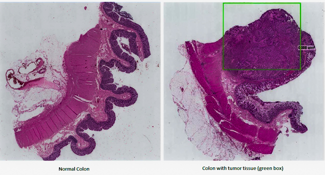
Artificial intelligence can be used as an image analysis tool to detect cancer tissue within tissue samples stained with hematoxylin and eosin (H&E). The cell nuclei, which are stained as purple, are of interest when performing a cancer diagnosis as cancer is often characterized by abnormal cell growth. Currently, pathologists perform cancer diagnoses by manually assessing the cellular tissue structures within large tissue samples, which may contain millions of cells. The manual assessment is subject to observer variability creating a risk that the patient does not receive the optimal treatment. Artificial intelligence has the potential to provide more robust diagnoses as the algorithms can be trained to recognize the appearance of different tissue structures. We have developed algorithms to recognize cancer tissue with the aim of making automated and more robust diagnoses in clinical practice. Current results have shown approx. 95% agreement between the algorithm and a pathologist in detecting cancer tissue. Additionally, the algorithm can be used to provide the pathologist with information about the location of the cancer tissue. The use of artificial intelligence therefore has the potential to improve the workflow within departments of pathology and provide patients with better diagnosis.